Fig. 3.

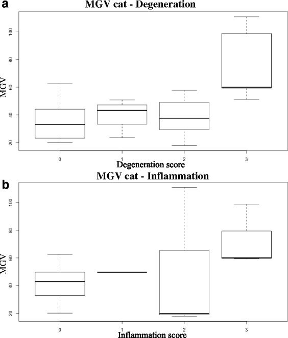
Box-and-whisker plots of MGV in the cat samples classified on the basis of the degeneration (a) and inflammation (b) scores assigned by the pathologists

Box-and-whisker plots of MGV in the cat samples classified on the basis of the degeneration (a) and inflammation (b) scores assigned by the pathologists