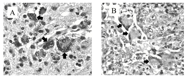
Figure 1.
Panel A. Hematoxylin and eosin (H & E) – stained thin section of hippocampus tissue from raccoon 01-2676. Syncytia are identified by large arrows. Some CDV inclusion bodies are indicated (small arrows). Original magnification × 200. Panel B. Thin section (H & E-stained) of lung tissue from raccoon 01-2663. Syncytia and CDV inclusion bodies are identified as in panel A.

