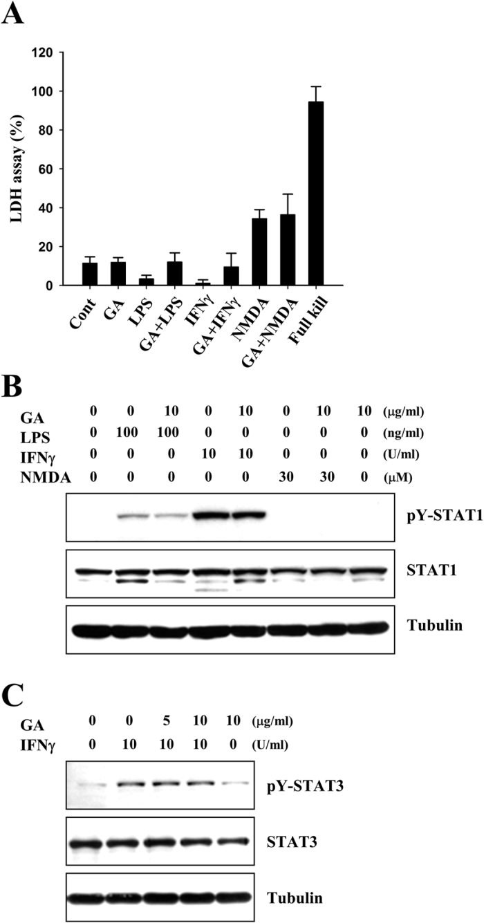
Figure 6. GA does not reduce the activations of STAT1 and −3 in neuronal cells.

(A) Rat primary cortical neurons were cultured and treated with 100 ng/ml LPS, 10 U/ml IFNγ, or 30 μM NMDA in the presence or absence of 10 μg/ml GA for 72 h. Bars represent the release of LDH from these primary cortical neurons. (B and C) Primary neuronal cells were mock-treated or treated with 10 μg/ml GA for 1 h, and then treated with 100 ng/ml LPS, 10 U/ml IFNγ, or 30 μM NMDA for 3 h. Western blot analyses were performed using the indicated antibodies. The results shown are representative of at least three individual experiments.
