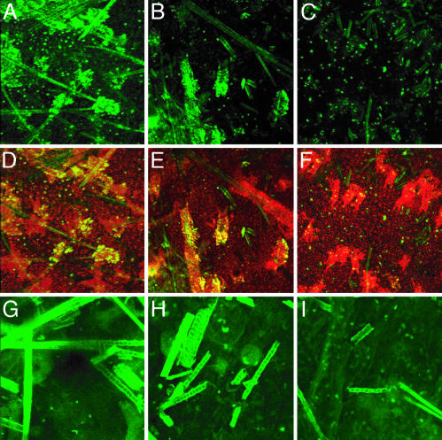
Fig. 4.
3D distribution of TUNEL-positive cells and active caspase-3-positive cells along hair shafts in epidermal sheets from pigmented mice irradiated with UVB. Mice were irradiated with 1,250 J/m2 UVB, and 24 h later epidermal sheets were prepared. Images were constructed by assembling confocal z axis sections. Shown are black (A, D, and G), yellow (B, E, and H), and albino (C, F, and I) strains. (A–C) TUNEL (green, fluorescein). (D–F) TUNEL merged with propidium iodide (red) and overlap in yellow. In pigmented mice exposed to UVB, TUNEL-positive cells were concentrated in the hair follicle and along the hair shafts. In the albino mouse (C and F), TUNEL-positive cells were distributed randomly and unrelated to the location of hair follicle. G–I show active caspase-3 only (green). Active caspase-3-positive cells were distributed randomly with respect to hair follicles in all irradiated genotypes. (Original magnification: A–F, ×100; G–I, ×200.)

