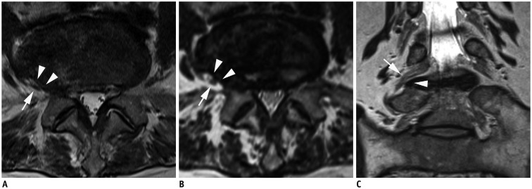
Fig. 3. MR images in 70-year-old woman with right radicular leg pain along L5 dermatome.
Axial 2D T2-weighted image (A) shows extraforaminal disc herniation and suspiciously deviated right L5 nerve root Axial 3D T2-weighted image (B) at same level shows suspiciously deviated right L5 nerve root having contact with herniated extraforaminal disc. Reader 1 missed this lesion on 2D images, and reader 2 missed this lesion on both 2D and 3D axial images. However, both readers correctly interpreted this lesion on 3D sequence with coronal and oblique coronal images. Superolaterally deviated right L5 nerve root is more definitely delineated on coronal 3D isotropic T2-weighted TSE image (C). There were no other findings compromising right L5 nerve root. MR = magnetic resonance, TSE = turbo spin-echo, 2D = two-dimensional, 3D = three-dimensional

