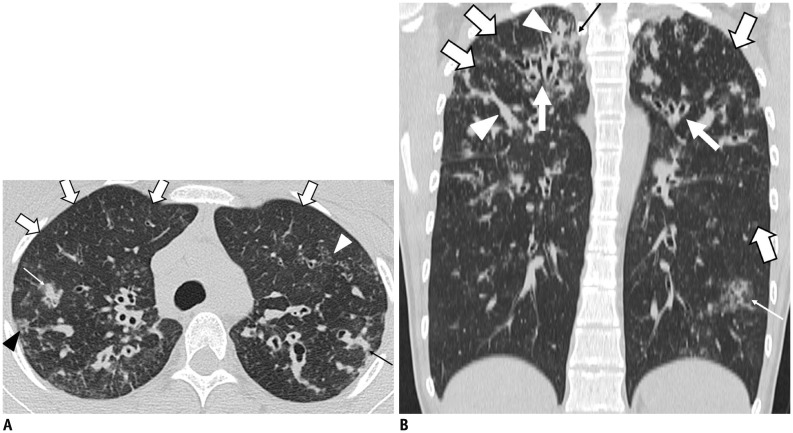
Fig. 3. 17-year-old female patient with cystic fibrosis and Pseudomonas aeruginosa infection.
A. Lung window image of thin-section (2.0-mm-section thickness) CT obtained at level of aortic arch shows extensive areas of bronchiectasis and cellular bronchiolitis (arrowheads) in both lungs. Also note wide areas (open arrows) of mosaic attenuation. Several areas of rectangular consolidation (thin arrows) suggest presence of concurrent bacterial pneumonia. B. Coronal reformatted (2.0-mm-section thickness) CT image demonstrates areas of bronchiectasis (arrows), mosaic attenuation (open arrows), and mucus plugging (arrowheads) predominantly involving bilateral upper lung zones. Also note areas of parenchymal opacity (thin arrows).

