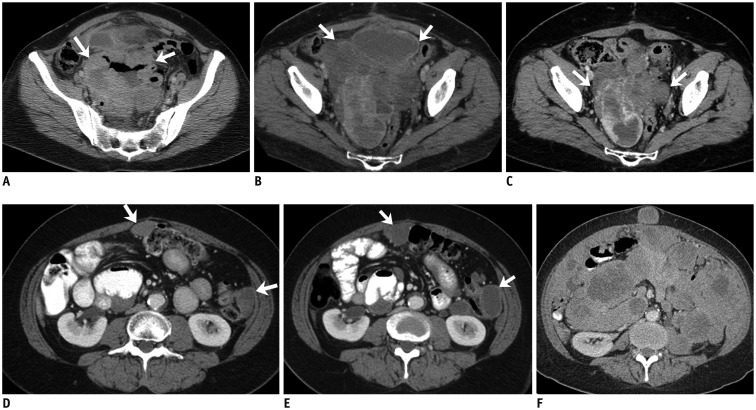
Fig. 2. 53-year-old woman with small bowel gastrointestinal stromal tumor with exon 9 mutation.
A. Axial contrast-enhanced CT image of lower abdomen reveals large cavitating mass in lower abdomen surrounded by small bowel loops (arrows). Patient underwent surgery which revealed small bowel mass. Histopathology revealed gastrointestinal stromal tumor with exon 9 mutation. B. Three months after surgery follow-up CT scan demonstrated recurrent pelvic mass (arrows). Patient was treated with high dose imatinib. C. CT scan after 6 months of treatment showed significant decrease in size of pelvic mass (arrows). Pelvic mass was excised and patient was restarted on high dose imatinib. D. Repeat CT scan after 4 months of treatment showed recurrence in form of multiple peritoneal masses (arrows). Patient was switched to sunitinib. E. CT scan performed 2 months after start of sunitinib therapy showed decrease in density of peritoneal deposits with mild increase in size (arrows). F. Follow-up CT scan three months later showed significant increase in peritoneal sarcomatosis. Patient died two months later. CT = computed tomography

