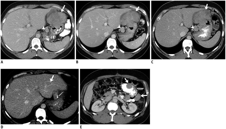
Fig. 3. 41-year-old man with gastric GIST with SDH mutation.
A. Axial contrast-enhanced CT image at time of diagnosis demonstrates large lobulated gastric mass (arrow). There is enlarged lymph node in gastrohepatic ligament (arrowhead). Patient was treated with neoadjuvant imatinib for one month. Follow-up CT scan showed no response to treatment and dose of imatinib was doubled. B. Repeat CT scan performed 3 months after therapy with high-dose imatinib showed no change in size of gastric mass and lymph node. Biopsy of mass at this time revealed SDH-deficient GIST. Patient was switched to sunitinib. C. CT scan after three months of treatment showed no response to treatment, instead new peritoneal nodule. Patient was taken up for surgery. D, E. Two years after surgery surveillance CT scan showed new liver metastasis (arrow, D) and peritoneal and bowel metastases (arrows, E). Patient was restarted on sunitinib. At time of last follow-up 7 years after initial diagnosis continues to have liver and peritoneal metastases which are stable in response to regorafenib therapy. CT = computed tomography, GIST = gastrointestinal stromal tumor, SDH = succinate dehydrogenase

