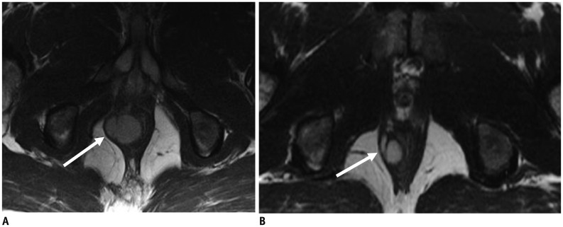
Fig. 4. 32-year-old man with anorectal gastrointestinal stromal tumor.
A. Axial T2-weighted MR image of pelvis reveals 3.3 cm mass (arrow) in anal canal. Biopsy of mass showed gastrointestinal stromal tumor with mitotic count of 1 per 50 high power fields. Tumor is low risk according to NIH consesus criteria, AFIP criteria and Joensuu criteria. Patient was treated with imatinib in neoadjuvant setting to downsize tumor. B. Follow-up MRI after 3 months of imatinib therapy shows decrease in size of mass (arrow). AFIP = Armed Forces Institute of Pathology, MRI = magnetic resonance imaging, NIH = National Institute of Health

