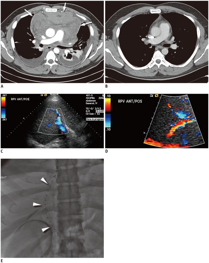
Fig. 10. Contrast-enhanced chest CT in patient with precursor T-cell lymphoma with surgical resection.
Baseline axial CT image (A) demonstrates large heterogeneously enhancing anterior mediastinal mass with associated pericardial effusion (arrows) and bilateral pleural effusions. Patient was taken for urgent pericardial window procedure, biopsy, which was initially thought to represent thymoma. Patient was treated with urgent radiation, chemotherapy with cytoxan, adriamycin, and cisplatin, and eventual surgical resection showing no residual disease in specimen, which was thought to be unusual for thymoma. Postsurgical axial CT image (B) demonstrates mild stranding in anterior mediastinum, consistent with postsurgical changes, without evidence for residual or recurrent lymphoma. Re-review of initial biopsy was felt to represent precursor T-cell lymphoma. Patient underwent induction chemotherapy per CALGB 9111 protocol and stem cell transplant. Following stem cell transplant, patient unfortunately developed veno-occlusive disease, as seen on ultrasound color Doppler images showing reversal of flow in right (C) and main (D) portal veins and expired despite transjugular intrahepatic portosystemic shunt procedure (arrowheads) (E).

