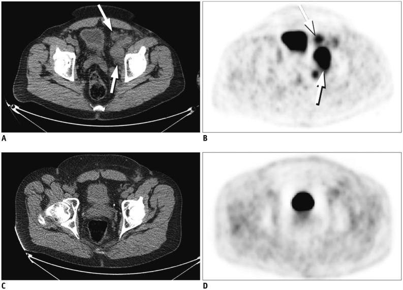
Fig. 5. 18FDG PET-CT in patient with anaplastic large cell lymphoma with treatment response.
Baseline CT (A) and PET (B) images demonstrate intense 18FDG uptake within left external iliac and pelvic sidewall lymphadenopathy (arrows). Post treatment CT (C) and PET (D) images demonstrate complete resolution of lymphadenopathy and 18FDG uptake after treatment with CHOP. FDG = flourodeoxyglucose, PET = positron emission tomography

