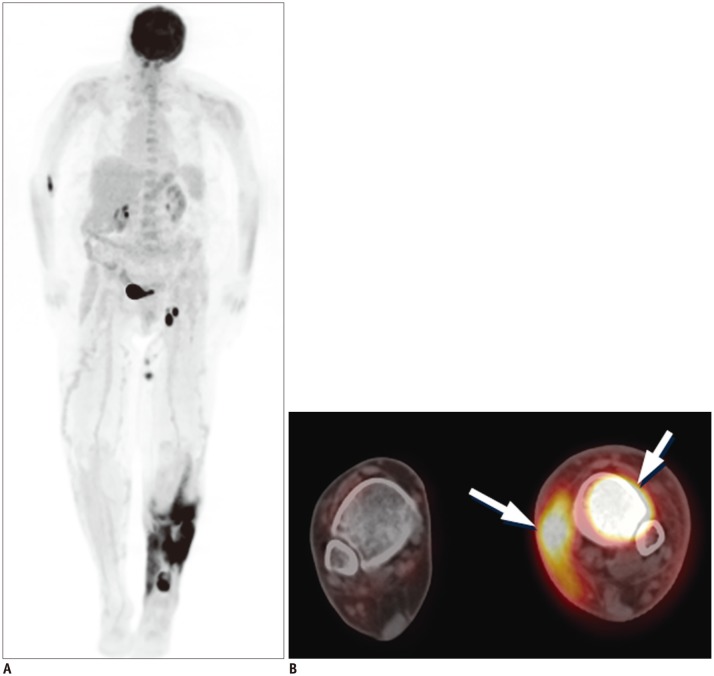
Fig. 7. 18FDG PET-CT in patient with extranodal natural killer T-cell lymphoma.
MIP (A) and axial fused PET-CT (B) images demonstrate intense 18FDG uptake in left lower extremity, involving marrow compartment of distal left tibia and surrounding subcutaneous soft tissue and overlying skin medially (arrows). FDG = flourodeoxyglucose, MIP = maximum intensity projection, PET = positron emission tomography

