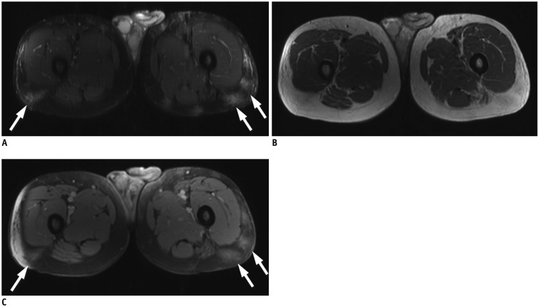
Fig. 8. Contrast-enhanced MRI in patient with subcutaneous panniculitis-like T-cell lymphoma.
Axial T2 fat-sat (A) and T1-weighted (B) images demonstrate vague areas of increased T2 hyperintensity and T1 isointensity in subcutaneous tissues of posterior proximal thighs (arrows). Axial T1-weighted postcontrast (C) image demonstrates corresponding areas of enhancement (arrows).

