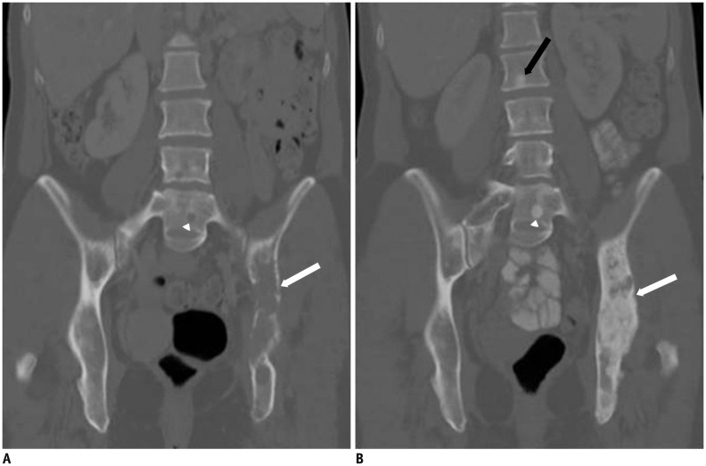
Fig. 3. 48-year-old woman with advanced breast cancer metastatic to bones.
A. Coronal CT image of the abdomen (bone window) reveal extensive lytic lesion involving left iliac bone (white arrow, A) and small lytic lesion within L5 vertebral body (white arrowhead, A). B. Follow-up coronal CT image of abdomen (bone window) shows marked increased sclerosis of left iliac lesion (white arrow, B), L5 vertebral body lesion (white arrowhead) and apparent new well-defined sclerotic lesion within L2 vertebral body (black arrow, B), which is consistent with treatment response.

