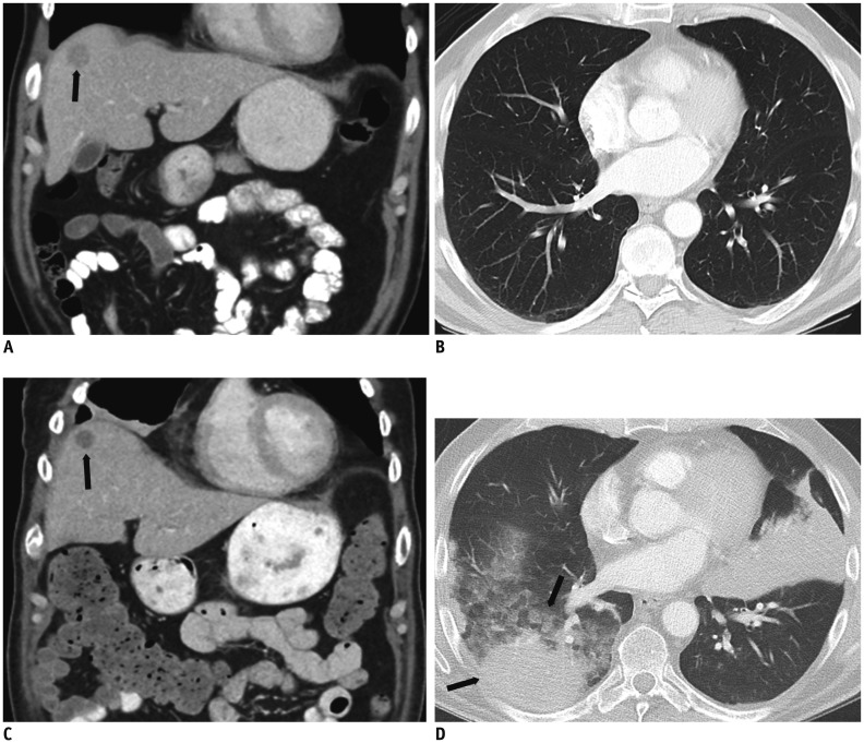
Fig. 1. 62-year-old male with history of ocular melanoma metastatic to liver.
A, B. Contrast enhanced coronal abdominal CT image shows single solid hepatic metastasis (black arrow, A); lung parenchyma is normal. C, D. After 8 weeks of treatment with combination therapy Ipilimumab/Nivolumab, patient presents to emergency department complaining of shortness of breath. While contrast enhanced coronal abdominal CT image shows decreased size and density of hepatic metastasis (black arrow, C), suggesting response to treatment, new lung consolidative opacities are concerning for drug pneumonitis (black arrows, D).

