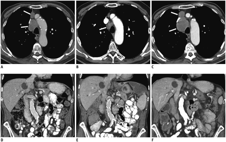
Fig. 3. 73-year-old gentleman with history of recurrent squamous cell lung cancer.
A, D. Axial contrast-enhanced CT image of mediastinum shows mediastinal adenopathy (white arrows, A); coronal abdominal image is unremarkable. B, E. After 4 weeks of treatment with Nivolumab, while mediastinal lymph nodes have significantly decreased (white arrow, B), visualized colon is hyperemic and fluid filled, suggesting colitis (black arrows, E). C, F. Nivolumab was stopped, high dose therapy with corticosteroid initiated. After 8 weeks, restaging contrast-enhanced abdominal CT shows resolution of colitis, but there has been significant increased in size of mediastinal adenopathy (white arrows, C).

