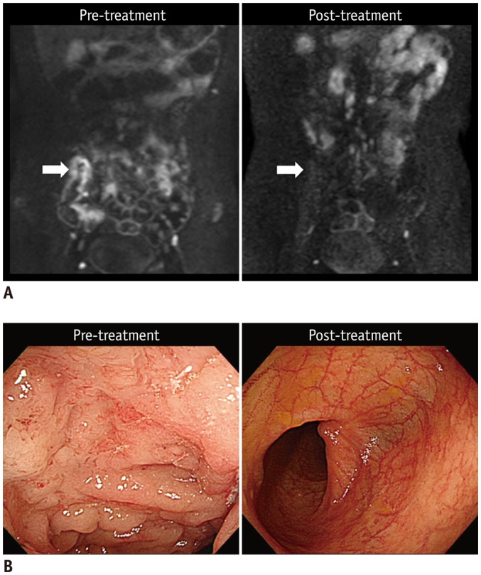
Fig. 3. 20-year-old man (at diagnosis) with CD who showed complete remission of terminal ileal lesion on endoscopy after 2 years of therapy.

A. Pre- and post-treatment DWI images (b = 900 s/mm2) indicate that restricted diffusion, which appears as hyperintensity in terminal ileum before treatment (left arrow), had completely disappeared at time of complete remission after treatment (right arrow). B. Colonoscopic images of terminal ileum shows complete resolution of multiple ulcers and mucosal swelling after treatment. CD = Crohn's disease, DWI = diffusion-weighted imaging
