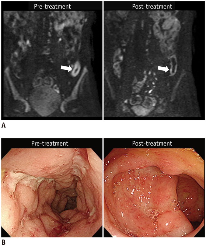
Fig. 4. 21-year-old man (at diagnosis) with CD shows reduced inflammation in descending colon on endoscopy after 1 year of therapy.

A. Pre- and post-treatment DWI images (b = 900 s/mm2) show that restricted diffusion, which appears as hyperintensity in distal descending colon, before treatment (left arrow) was remarkably reduced after treatment (right arrow). B. Colonoscopic images of corresponding area show large deep longitudinal ulcers before treatment (left), and reduced inflammation after treatment, presented as scattered small superficial ulcers and aphthoid lesions (right). CD = Crohn's disease, DWI = diffusion-weighted imaging
