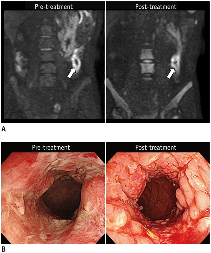
Fig. 5. 25-year-old man (at diagnosis) with CD who showed unchanged inflammation in descending colon on endoscopy after 2 years of therapy.

A. Pre- and post-treatment DWI images (b = 900 s/mm2) reveal similar restricted diffusion that appears as hyperintensity in descending colon (arrows). B. Colonoscopic images of corresponding area reveal persistent extensive inflammation with multiple large ulcers before treatment and on follow-up without remarkable improvement. CD = Crohn's disease, DWI = diffusion-weighted imaging
