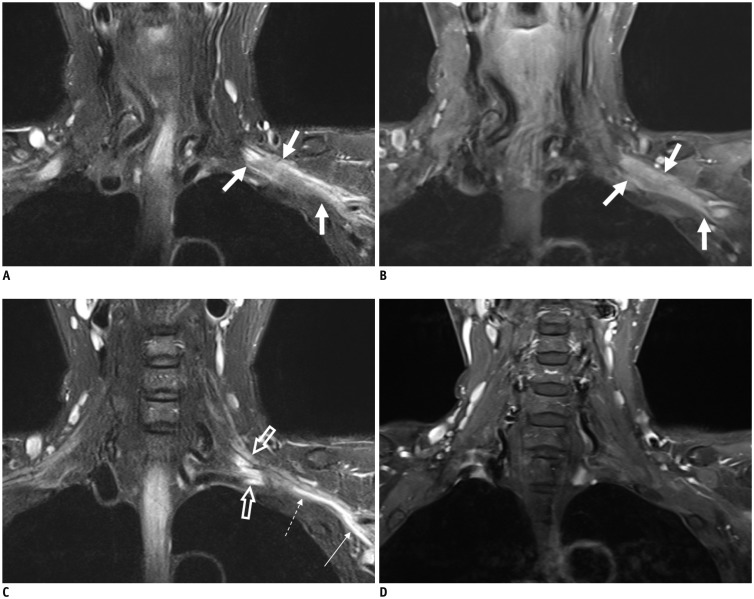
Fig. 5. 58-year-old female with left breast cancer.
Coronal fat-suppressed T2-weighted images (FS T2WIs) (A) and contrast-enhanced (CE) FS T1-weighted image (T1WI) (B) showing infiltrative and enhanced tumor involving entire trunks of left brachial plexus (arrows). Coronal FS T2WI (C) depicts hyperintense change without enhancement of roots (empty arrows), part of lower trunk (dotted arrow), and medial cord (thin arrow) originating from secondary compressive plexopathy, different from infiltrative tumor (thick arrows on A and B). There is no enhancement at corresponding segments of left brachial plexus on coronal CE FS T1WI (D).

