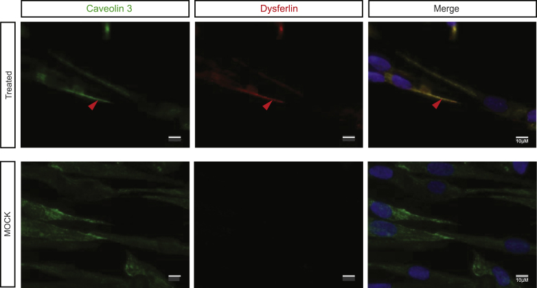
Fig.2.
Exon 32-skipped dysferlin is correctly localized. Caveolin 3 labeling: maturation of myotubes and production of dysferlin were evidenced by Hamlet 1 labeling (Bars = 10 μm, arrow pointed to colocalized signal). DAPI was used as a nucleus marker. All images were captured by an apotome microscope.

