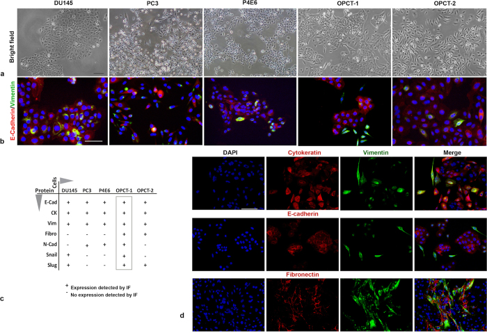
Figure 2. Identification of OPCT-1 as a suitable model for the study of spontaneous EMT in prostate cancer.
(a) Bright field images of human prostate cancer cell lines derived from metastatic lesions: DU145 and PC3, and primary tissues: P4E6, OPCT-1 and OPCT-2 (Image magnification at x10). (b) Dual immunofluorescent staining of DU145, PC3, P4E6, OPCT-1 and OPCT-2 using antibodies against E-cadherin (red) and vimentin (green) (n = 3). (c) Table summarising the results of the IF screening of DU145, PC3, P4E6, OPCT-1 and OPCT-2 cells for the expression of several EMT-associated markers. (d) Summary composite of OPCT-1 stained with common markers used to investigate EMT: Cytokeratin pan/vimentin, E-cadherin/vimentin, fibronectin/vimentin. Scale bar: 50 μM.

