Figure 7.
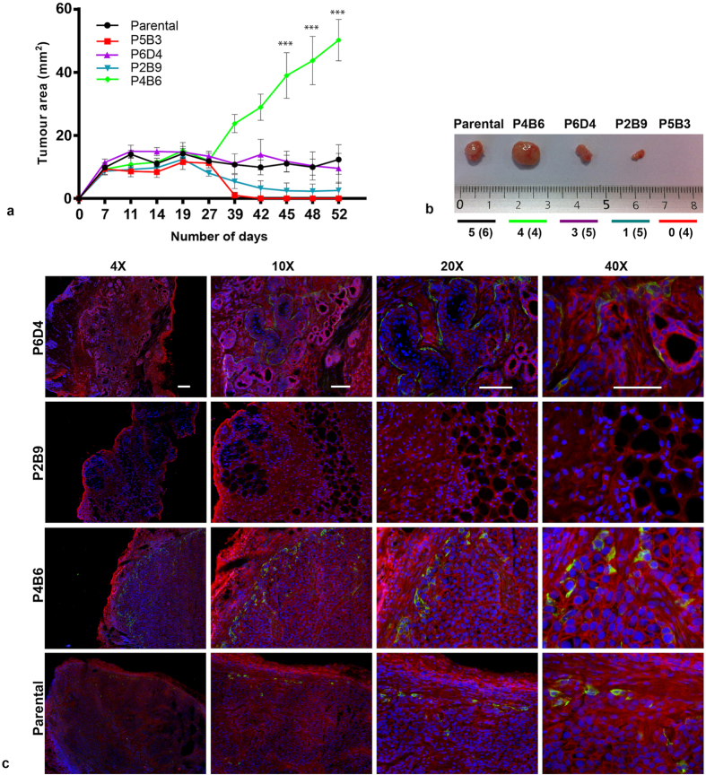
(a) In vivo tumourigenesis assay. Clones P5B3, P6D4, P2B9, P4B6 and parental OPCT-1 were injected subcutaneously into the right flanks of male athymic nude mice (6 animals per cell line). Tumour growth was monitored using calliper measurements and mice were euthanised once one of the tumours reached 1 cm in diameter. Data are presented as the mean tumour area ± SD. Statistical significance was calculated using the Student’s t-test (significance indicated as asterisks). (b) Representative tumours excised from tumour-bearing mice arranged in descending order of the clones’ in vitro vimentin positivity. (c) Immunofluorescent staining of tumour sections derived from clones P5B3, P6D4, P2B9, P4B6 and parental OPCT-1 for E-cadherin (red) and vimentin (green) expression. Representative images. (×10, ×20 and ×40 magnification, n = 3). Scale bar: 50 μM.

