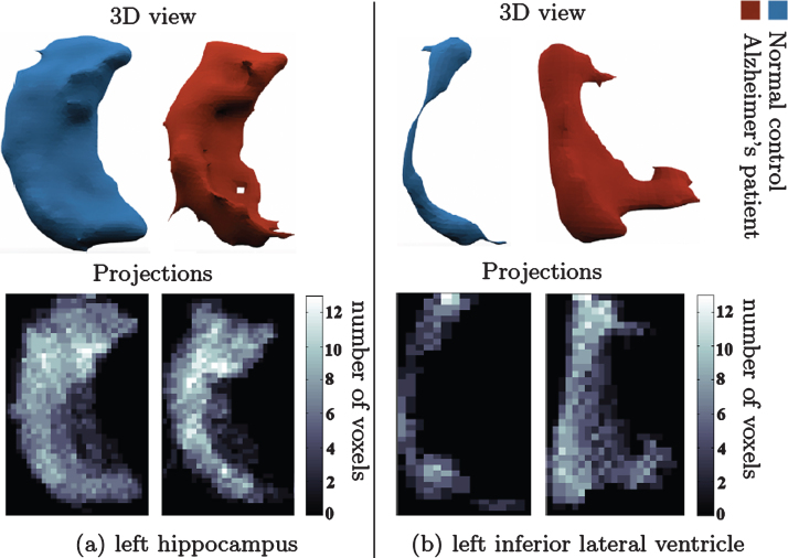
Fig.2.
Change in shape of brain structures in AD manifested on projection images. (a) l. hippocampus 3D shape (upper panel) in NC (left) and AD (right). (b) l. inf. lat. ventricle 3D shape (upper panel) in healthy control (left) and AD patient (right). Lower panel shows the corresponding canonical view using our principal projections method.

