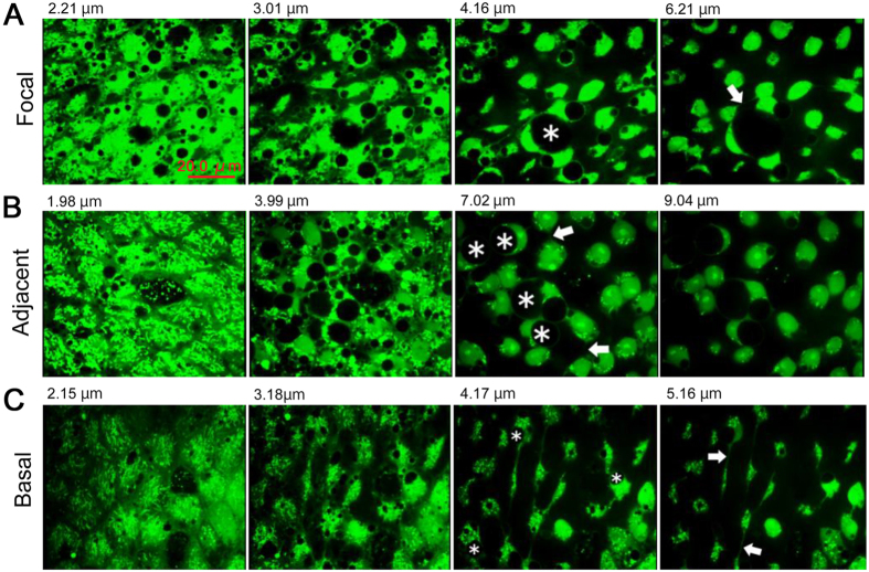
Figure 6. Serial optical sections of epithelial cells stained with CFSE.
Panels are presented from the apical surface (left) to deeper layers (right). Depth from the apical surface is indicated in each panel. Endosome-like structures (asterisks) and horizontal processes that connect two cells (arrows) are indicated. (A) Focal region. See also Supplementary Video 3. (B) Adjacent region. See also Supplementary Video 4. (C) Basal region. See also Supplementary Video 5.

