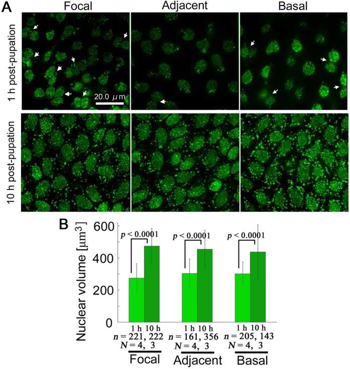
Figure 7. Comparison between 1 h and 10 h post-pupation.
(A) SYBR Green I staining for nuclei. Two (or more) nuclei are associated together, especially in the focal region (arrows) 1 h post-pupation. Larger nuclei are densely packed at 10 h post-pupation. (B) Changes in nuclear volume. The number of cells examined (n) and the number of pupae used (N) are indicated.

