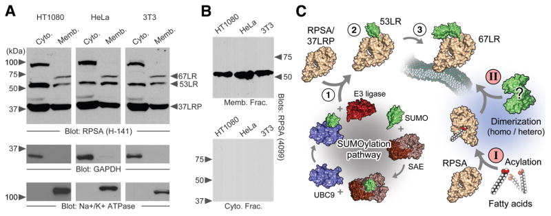
Figure 1.
The higher molecular weight species of RPSA. A – SDS-PAGE of cytosolic and membrane extracts from HT1080, HeLa and NIH 3T3 cells. The monomeric 37-kDa RPSA is indicated along with higher molecular weight species. GAPDH and the Na+/K+-ATPase are shown as cytosolic and membrane markers, respectively. H-141 antibody is used to detect RPSA. B – As above with RPSA 4099-1 antibody. C – Two hypothetical pathways leading to the construction of 67LR from a 37-kDa RPSA precursor (37LRP). Progressive SUMOylation (1-2-3) and fatty acid acylation (I) followed by either a homo- or heterodimerization event (II) are depicted.

