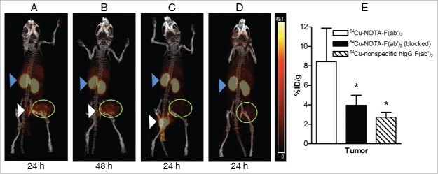
Figure 3.
Whole-body microPET/CT images of mice with subcutaneous SK-OV-3 HER2-overexpressing human ovarian cancer xenografts at 24 h post-injection (p.i.) (A) or 48 h p.i. (B) of 64Cu-NOTA-pertuzumab F(ab')2. (C) Images obtained at 24 h p.i. of 64Cu-NOTA-pertuzumab F(ab')2 with pre-administration of 1 mg of pertuzumab 24 h prior to radiopharmaceutical injection. (D) Images obtained at 24 h p.i. with 64Cu-labeled nonspecific hIgG F(ab')2. Tumor xenografts are indicated by the green circle. Also visualized are the kidneys (blue arrowhead) and bladder/urine (white arrowhead). The HER2 specificity of tumor uptake of 64Cu-NOTA-pertuzumab F(ab')2 was confirmed by biodistribution studies at 48 h p.i. (E) showing a significant decrease in tumor uptake of the radiopharmaceutical in mice pre-administered excess unlabeled pertuzumab to block HER2 or injected with 64Cu-labeled non-specific hIgG F(ab')2 (*P < 0.05).

