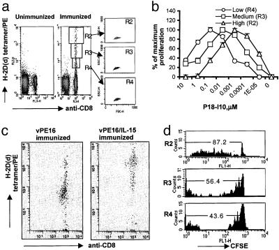
Fig. 2.
In vivo proliferation and persistence of antigen-specific CD8+ T cells depend on CD8+ T cell avidity. (a) Two to 3 months after boosting, splenocytes were stained with anti-CD8 and P18-I10 tetramer concurrently, as described in Materials and Methods. Cells at Left were from unimmunized animals. The three graphs at Right show the distribution of cells from gate R2, R3, and R4 in forward and side scatter plots. (b) Cells were gated, based on the brightness of tetramer staining, and sorted, left in the media for 6 h, and stimulated with P18-I10-pulsed splenocytes. Proliferation was measured as in Fig. 1. (c) Four months after boosting, spleen CD8+ T cells from mice immunized with vPE16 (Left) or vPE16/IL-15 (Right) were stained with anti-CD8 and P18-I10 tetramer, and then double-positive cells were sorted and reanalyzed by flow cytometry. (d) Two to 3 months after the boost, spleen CD8+ T cells from the immunized mice were labeled with CFSE and transferred to naïve animals. Four to 5 weeks after the transfer, spleen cells in the recipients were stained with anti-CD8 and tetramer, gated as in a, and analyzed to measure homeostatic proliferation. The numbers in histograms are mean ± SEM of at least four individual recipients in each experiment. Three repeated experiments showed consistent results.

