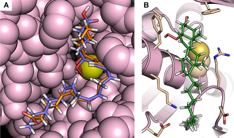
Fig. S1.
Representative docking poses. (A) Representative docking poses of 5(S)-HpETE and 15(S)-HpETE in the XfOhr active site showing great similarity in the overall conformation of the hydrocarbon chains of these arachidonic acid derivatives. In both cases, the hydroperoxide groups are positioned close to Cys61 (yellow), although their carboxyl groups are positioned on opposite sides of the active site. The atoms belonging to the enzyme are represented as spheres and colored in light pink. The 5(S)-HpETE is shown in blue, and the 15(S)-HpETE is shown in orange. (B) Docking of cholesterol-5α-hydroperoxide in the XfOhr active site. Note the hydroperoxide group faces away from Cys61 (yellow), whose side chain is represented as spheres. XfOhr is colored in light pink and represented as a cartoon. Residues within 4 Å of the cholesterol-5α-hydroperoxide molecule are colored in beige and represented as sticks, whereas cholesterol-5α-hydroperoxide itself is colored in green.

