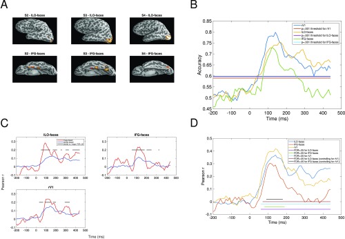
Fig. S2.
Analyses for left hemisphere. (A) Face-selective regions in left hemisphere, for each participant in which regions could be identified. (B) Classification accuracy as a function of time (milliseconds) and region (lV1, lLO-faces, and lFG-faces). All other details as in Fig. 4. (C) Correlations between neural data and image-based and identity-based representations of our stimuli, as a function of time (milliseconds). All other details as in Fig. 5. (D) Correlations between behavioral and neural data, as a function of time (milliseconds). All other details as in Fig. 6B.

