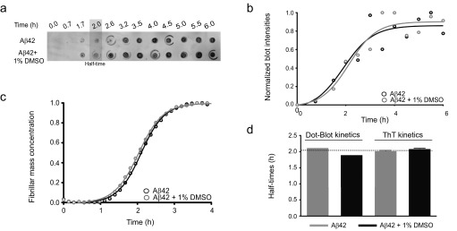
Fig. S1.
Characterization of the effects of DMSO on Aβ42 aggregation by ThT-based kinetics and immunochemistry. (A) Time course of the formation of 2 μM Aβ42 fibrils as assessed by the fibril-specific OC antibody in the absence and the presence of 1% DMSO. The OC antibody probes fibrillar structures that have started to form roughly around 2 h. (B) Quantitative time course showing the evolution of the fibril formation of 2 μM Aβ42 based on the intensities of the dot-blot assay from A in the absence and presence of 1% DMSO. (C) ThT-based kinetics of 2 μM Aβ42 in the absence and presence of 1% DMSO as a function of time. (D) Comparative analysis between the half-times derived from either the dot-blot intensities (B) or the ThT-based kinetics (C) showing similar values (i.e., 2 h), and thus excluding an effect of 1% DMSO on the aggregation kinetics.

