Fig. S5.
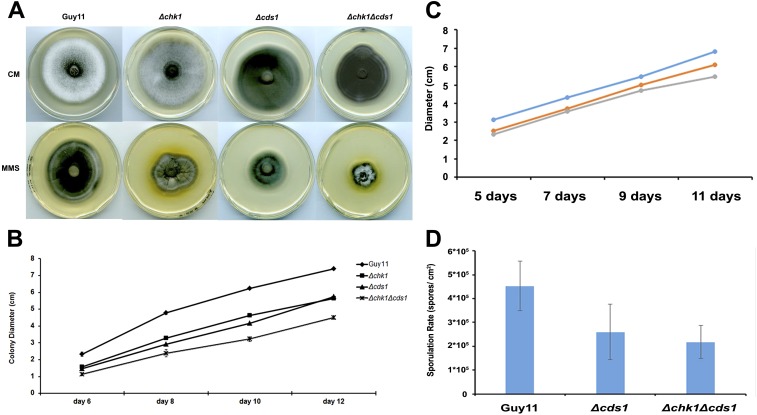
Vegetative growth and colony morphology of DNA replication checkpoint mutants. (A) Mycelial plugs of Guy11, Δchk1, Δcds1, and Δchk1Δcds1 mutants, respectively, were inoculated onto 0.001% of the alkylating agent methyl methanesulfonate (MMS) and in CM plates and incubated during 12 d at 26 °C. Images were taken by using an Epson Expression 1680 Pro scanner after 10 d postinoculation. (B) Colony growth of Guy11, Δchk1, Δcds1, and Δchk1Δcds1 over the period of 12 d. Error bars represented the SE from three independent replicates. (C) Colony growth of Guy11, Δchk1, Δcds1, and Δchk1Δcds1 mutants during a period of 12-d incubation in complete medium. Error bars represent the SE from three independent replicates. (D) Bar chart to show sporulation rate (spores per cm2) of Guy11, Δcds1, and Δchk1Δcds1. Error bars represented the SE from three independent replicates.

