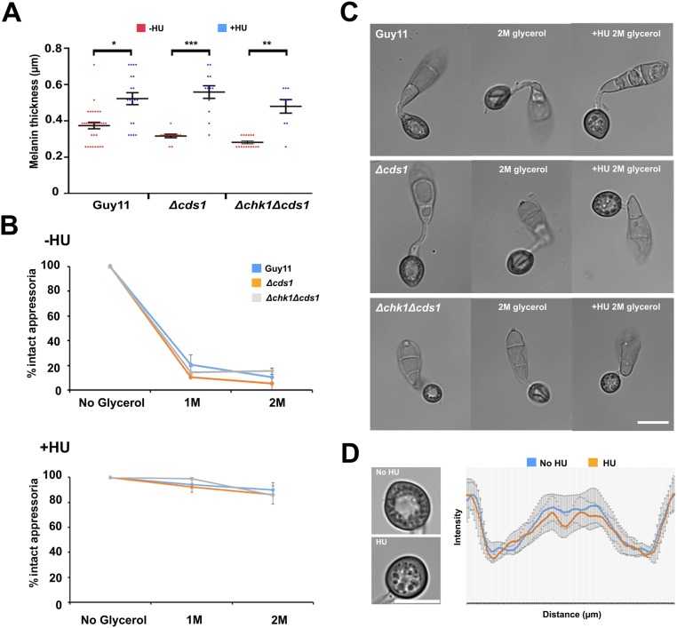
Fig. S10.
Arrest in S phase increases melanin content and turgor generation of appressoria, independent of the DDR. (A) Arrest at S phase increases the thickness of the melanin layer, independently of the DDR. Scatter dot plot to compare melanin layer thickness after exposure to HU. Appressoria were allowed to form on hydrophobic plastic coverslips for 10 h, when 200 mM HU was added. At 24 h, micrographs were obtained to quantify the melanin layer thickness by measuring the intensity profile values over line scan diameters that crossed the center of the appressorium. *P < 0.05; **P < 0.01; ***P < 0.001 (nonparametric Kruskal–Wallis test; n = 2 experiments; appressoria observed = 8–30). (B) Micrographs to show the effect of exposure to 2 M glycerol at 24 h, after addition of HU at 10 hpi in Guy11, Δcds1, and Δchk1Δcds1 mutants. (C) Incipient cytorrhysis assay to measure appressorium turgor generation in Guy11, Δcds1, and Δchk1Δcds1 mutants in the presence or absence of HU exposure (added at 10 hpi). Appressoria were allowed to form on hydrophobic plastic coverslips for 10 h, when 200 mM HU was added. At 24 h, appressoria were exposed to 1 and 2 M of glycerol concentration, and the percentage of intact appressoria was recorded (n = 2 experiments; appressoria observed = 84–134). (D) The pore was no longer formed in the presence of HU. Appressoria were allowed to form on hydrophobic plastic coverslips for 10 h, when 200 mM HU was added. At 24 h, micrographs were obtained to show absence of the appressorium pore when the inhibitor was added (n = 3 experiments; appressoria observed = 45). The graph shows the mean of intensity profile values overlaid (±SE) over 12-µm linescan diameter that crosses the appressorium through the center. (Scale bars, 10 μm.)

