Figure 6.
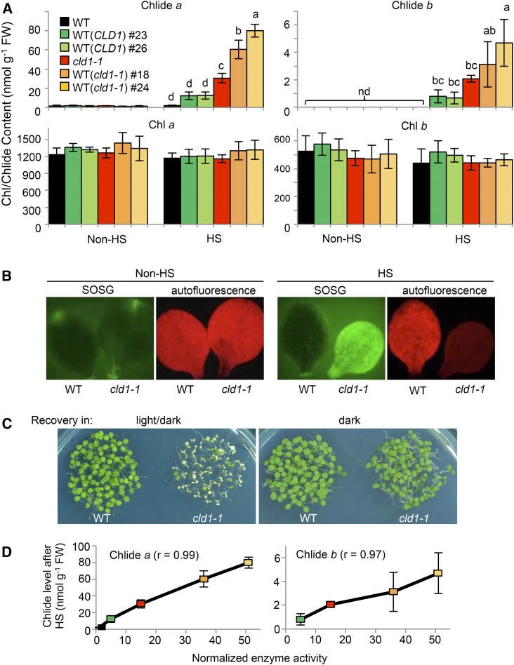
Heat-Induced Accumulation of Chlide a and b Is Associated with Supraoptimal CLD1 Activities, Resulting in Singlet Oxygen Formation and Light-Dependent Cotyledon Bleaching.
(A) Levels of Chlides and chlorophylls extracted from pooled 5-d-old seedlings before and after treatment at 40°C for 1 h (HS). The line designations are as described in Figure 1. Data are means ± sd of three independent experiments. One-way ANOVA with Tukey’s method was applied to show the significant differences at P < 0.01 in pairwise comparison and classified with the letter a, b, c, or d. ANOVA tables can be found in Supplemental Figure 10. Column without any letter indicates no significant difference from that of the wild type before HS.
(B) Detection of singlet oxygen in wild-type and cld1-1 seedlings. After HS, the singlet oxygen level was analyzed by incubating the cut cotyledons with 10 μM SOSG solution under light for 4 h at 22°C. Non-heat-treated samples were used as the control. Images with false color were obtained under a fluorescence microscope. Green indicates the accumulation of singlet oxygen, and red indicates the autofluorescence of chlorophyll.
(C) Heat-induced cotyledon bleaching in cld1-1 is light-dependent. Five-d-old seedlings were treated at 40°C for 1 h and recovered for 2 d at 22°C under 16-h/8-h light-dark cycle (100 μmole m−2 s−1) and dark. The pictures were obtained right after recovery.
(D) Correlation of levels of heat-induced Chlide a and b versus the normalized chlorophyll dephytylase activities in indicated lines. The enzyme activity in each line was calculated by multiplying the protein level shown in Figure 3A and the normalized reaction rate determined in Figure 5B. The activity in the wild type is assigned to 1. The color codes are as in (A). Pearson’s correlation coefficients (R) are shown.

