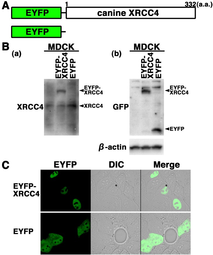
Fig. 3.
Subcellular localization of EYFP-canine XRCC4 in living canine cells. (A) Schematics of EYFP-canine XRCC4 chimeric protein and control protein (EYFP). (B) EYFP-canine XRCC4 was expressed in canine (MDCK) cells, and the expression of EYFP-canine XRCC4 was examined by Western blotting using the anti-XRCC4, anti-GFP or anti-β-actin antibody. (C) Imaging of living EYFP-canine XRCC4-transfected cells. Living MDCK cells transiently expressing EYFP-canine XRCC4 or EYFP were analyzed by confocal laser microscopy. EYFP images for the same cells are shown alone (left panel) or merged (right panel) with differential interference contrast images (DIC) (center panel).

