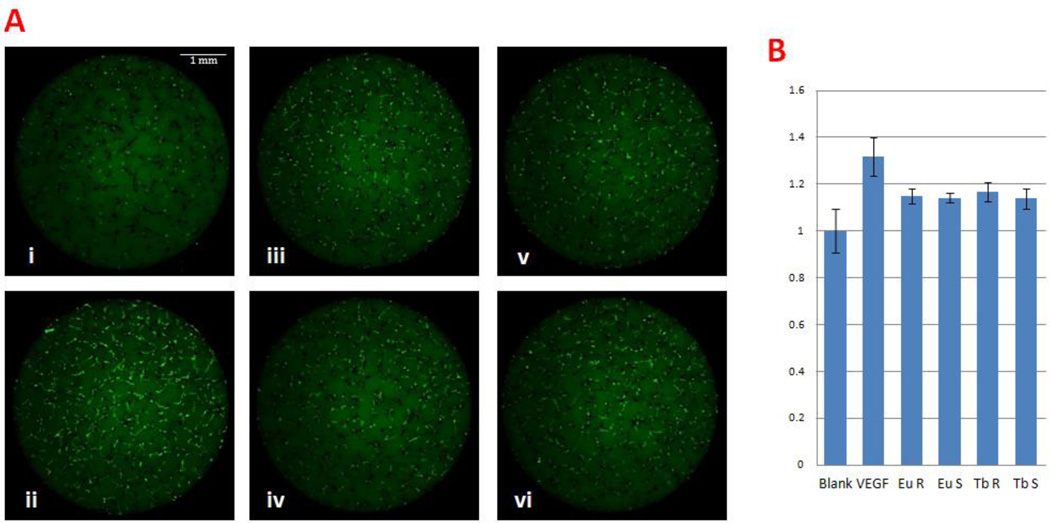
Figure 3.
Lanthanide nanoparticles induced angiogenesis in vitro. A. Embryonic primary cell culture with 1µg/ml nanoparticles or 20 ng/ml VEGF. i) Blank control, ii) 20 ng/ml VEGF, iii) 1 µg/ml Eu Rods, iv) 1 µg/ml Eu Spheres, v) 1 µg/ml Tb Rods, vi) 1 µg/ml Tb Spheres. VEGF exerted significantly improved GFP+ cell proliferation. Nanoparticles also show proangiogenesis ability. B. Quantitative analysis by Meta Express High Content Screening software.

