Figure 4.
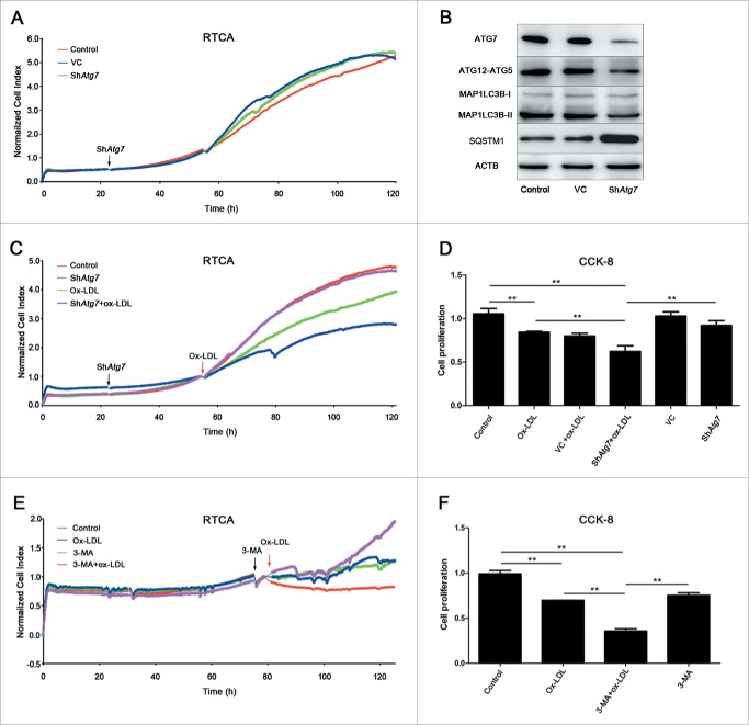
Autophagy was a protective response in EPC proliferation. (A) EPCs were seeded on E-plates for 24 h before lentiviral vector containing shRNA targeting Atg7 (shAtg7) infection (black arrow). RTCA indicated no significant difference among control, vector-control (VC) and shAtg7 groups. (B) Representative western blots for the detection of ATG7, ATG12–ATG5 conjugate, MAP1LC3B-II and SQSTM1 after infection showed that Atg7 was successfully knocked down and autophagy was effectively inhibited in Atg7 silencing group. (C) Cells were exposed to ox-LDL (60 μg/ml) (red arrow) after shAtg7 infection. The normalized cell index indicated that EPC proliferative activity fell down more quickly than ox-LDL alone and control groups after silencing Atg7. (D) CCK-8 assay showed a similar result that silencing Atg7 before ox-LDL treatment significantly reduced proliferative activity compared with ox-LDL alone and control groups. (E) 3-MA (2 mM) was added to inhibit autophagy (black arrow) before 60 μg/ml ox-LDL (red arrow). The normalized cell index indicated that after 3-MA inhibition, EPC proliferative activity fell down more quickly than ox-LDL alone and control groups. (F) CCK-8 assay showed similar results that the proliferative activity in 3-MA+ox-LDL group reduced significantly compared with ox-LDL alone and control groups. (Cells were isolated from 3 rats for one experiment and 3 independent experiments were performed, mean + SD, **P < 0.01).

