Fig. 3.
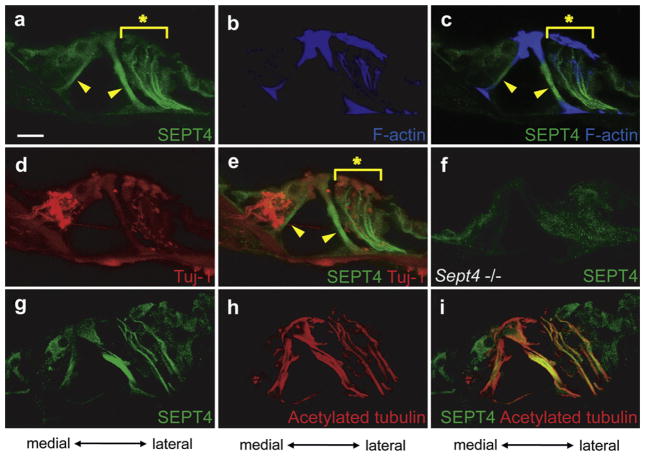
Expression pattern of SEPT4 in the adult mouse cochlea. a–e. Immunohistochemistry of the wild type adult mouse cochlea from a same section. SEPT4 (a), F-actin (b), SEPT4 and F-actin (c), β3-tubulin (Tuj-1) (d), and SEPT4 and β3-tubulin (Tuj-1) (e) were detected. f. Immunohistochemistry of adult Sept4 null mice cochlea for SEPT4. g–i. Immunohistochemistry of the wild type adult mouse cochlea from a same section. SEPT4 (g), acetylated α tubulin (h), and SEPT4 and acetylated α tubulin (i) were detected. SEPT4 (green) was strongly expressed in pillar cells (Arrowheads in a, c, and e). SEPT4 was also strongly expressed in filament-like structures in the lateral side of the organ of Corti from the basilar membrane to the reticular lamina (Asterisks in a, c, and e), presumably in the Deiters’ cells. SEPT4 signal overlapped with that of F-actin (blue) (b and c) and acetylated α tubulin (red) (h and i), but did not overlap with that of Tuj-1 (red) that is expressed in nerve fibers (d and e). These data indicated that SEPT4 was expressed in Deiters’ cells. SEPT4 was also expressed in hair cells and other supporting cells in weaker intensity. Detection of SEPT4 antibodies in Sept4 null mice cochlea was served as negative control of these experiments (f). Scale bar in a indicates 15 μm. (For interpretation of the references to color in this figure legend, the reader is referred to the web version of this article.)

