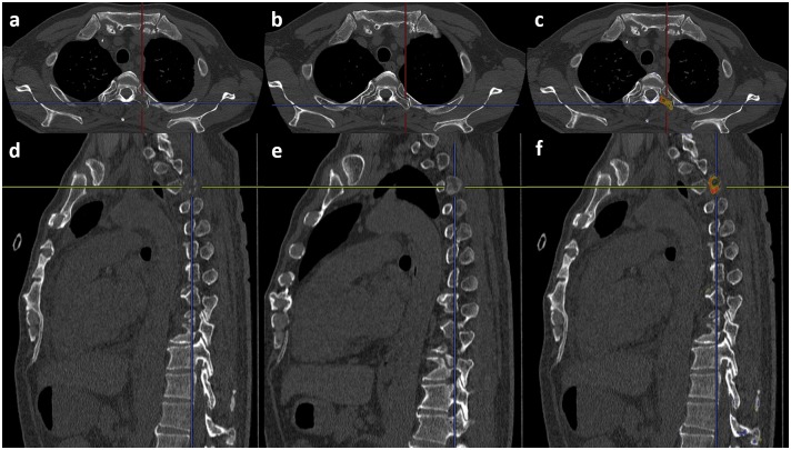
Fig 3. A case of osteolytic metastasis in the left transverse process of the third thoracic vertebra.
(a) Follow-up axial image. (b) Initial axial image. (c) Result of the 3D-CT subtraction. The yellow and red colors indicate the metastasis. (d) Follow-up sagittal image. (e) Initial sagittal image. (f) Result of the 3D-CT subtraction. The yellow and red colors indicate the metastasis.

