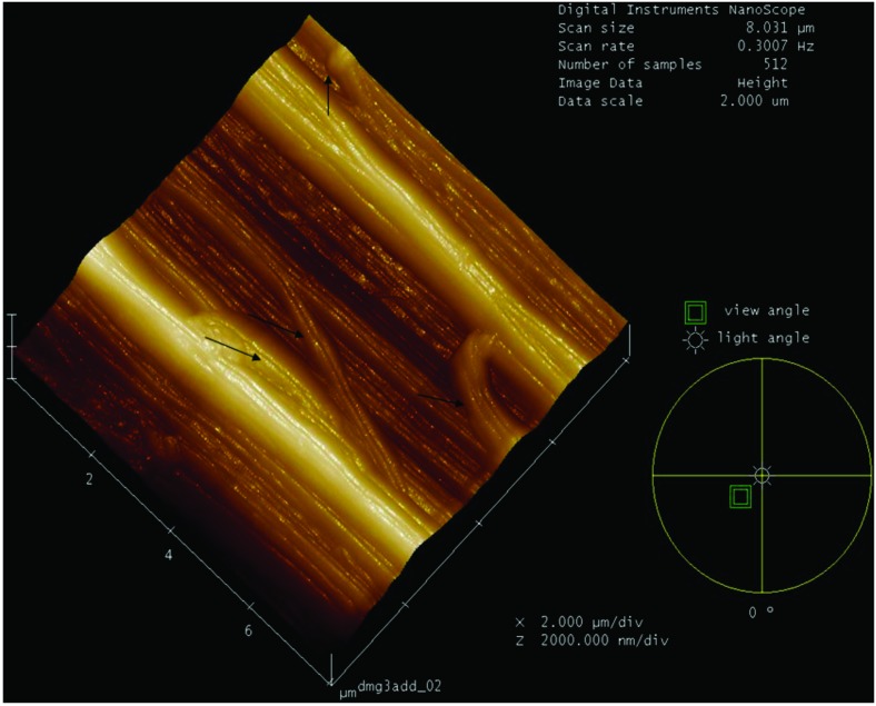
Fig 4. 3D representation of the surface topography of the fibrils of the Achilles tendon of a Diabetic Group animal, showing the change in direction of the collagen fibers.
The fibers were displaced in opposite directions or exhibited bifurcations, resulting in loss of morphologic and collagen nano-structure features, with changes in the direction of the collagen fibrils indicated by arrows.

