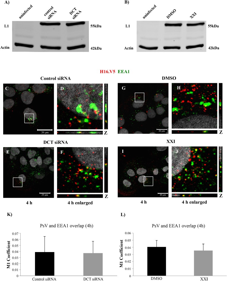
Fig 2. DCT siRNA or XXI treatment does not interfere with binding and early endosome localization of HPV16 PsV in HaCaTs.
Western blot analysis of L1 and actin levels in (A) uninfected, control and DCT siRNA transfected HaCaTs, (B) uninfected, DMSO and 300pM XXI treated HaCaTs. (C-J) Immunofluorescence analyses of EEA1 (green), L1 (red) in control and DCT siRNA transfected and DMSO and XXI treated cells. Nuclei are stained with DAPI (grey). Colocalization of L1 and EEA1 appears yellow. (K and L) The JACoP plugin for ImageJ was used to measure the M1 coefficient (fraction of red signal overlapping with green signal) with three confocal scans for each condition.

