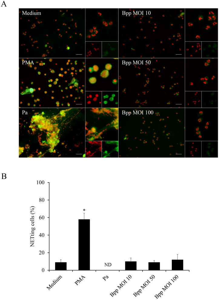
Fig 3. B. parapertussis precludes NETs release in resting neutrophils.
Neutrophils were infected with non-opsonized B. parapertussis (Bpp) (MOI: 10, 50 or 100) or P. aeruginosa (Pa) (MOI: 10) for 4 h at 37°C. Samples were fixed and permeabilized prior to labeling the NE in green and neutrophil DNA with a red fluorescent dye. Neutrophils incubated with PMA or medium alone were used as a positive and negative control, respectively. Samples were analyzed by fluorescence microscopy and the number of NETting cells was determined by the ImageJ softaware. At least 500 cells were counted per sample. (A) Representative microscopy images of neutrophils 4 h post infection are shown. Scale bar: 20 μm. (B) The bars represent the percentage of neutrophils that underwent NETosis. The data represent the mean ± SD of three experiments with neutrophils from different donors. ND: non-determinable (NETotic cells are no longer observed; only big clusters of NETs are seen). The percentage of NETting cells in PMA treated neutrophils was significantly different from the percentage of NETting cells observed in the other samples (*P <0.05).

