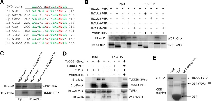
Fig 3. WDR1 forms a complex with TbCUL4 and TbDDB1.
(A). Alignment of the putative DWD box in WDR1 with the DWD box of fission yeast and human WD40-repeat proteins that have been confirmed to bind to DDB1. The three highly conserved residues are highlighted in red, and other conserved residues are in green. The consensus sequence of the DWD box is shown at the top of the aligned sequences. Tb, T. brucei; Sp, Schizosaccharomyces pombe; Hs, Homo sapiens. (B). WDR1 interacts with TbCUL4 but not other Cullin proteins, in T. brucei, as demonstrated by co-immunoprecipitation. WDR1-3HA and each of the five PTP-tagged Cullin proteins were co-expressed from their respective endogenous locus in T. brucei. Immunoprecipitation was performed by incubating the cell lysate with IgG beads, and immunoprecipitated proteins were then immunoblotted with anti-HA antibody and anti-Protein A (α-ProtA) antibody, respectively. (C). WDR1 interacts with TbDDB1 in vivo in T. brucei, as demonstrated by co-immunoprecipitation. WDR1-3HA and TbDDB1-PTP were co-expressed in T. brucei, and immunoprecipitation and Western blotting were performed as described in panel B. (D). WDR1, TbCUL4, TbDDB1 and TbPLK form a complex in T. brucei, as demonstrated by co-immunoprecipitation. WDR1-3HA, TbCUL4-PTP and TbDDB1-3Myc were co-expressed from their respective endogenous locus in T. brucei. Immunoprecipitation of WDR1-3HA was carried out by incubating the cell lysate with EZview Red anti-HA affinity gel, and immunoprecipitated proteins were immunoblotted with anti-HA antibody, anti-Myc antibody, anti-TbPLK antibody and anti-Protein A (α-ProtA) antibody to detect WDR1-3HA, TbDDB1-3Myc, TbPLK and TbCUL4-PTP, respectively. (E). The N-terminal domain of WDR1 mediates the interaction with TbDDB1, as demonstrated by in vitro GST pull-down. The N-terminal fragment (1–300 aa) of WDR1, which contains the WD40 motif, was expressed as a GST-fusion protein in E. coli, purified and used to pull down TbDDB1-3HA from T. brucei cell lysate. Purified GST-WDR11-300 and GST were stained by coomassie brilliant blue (CBB).

