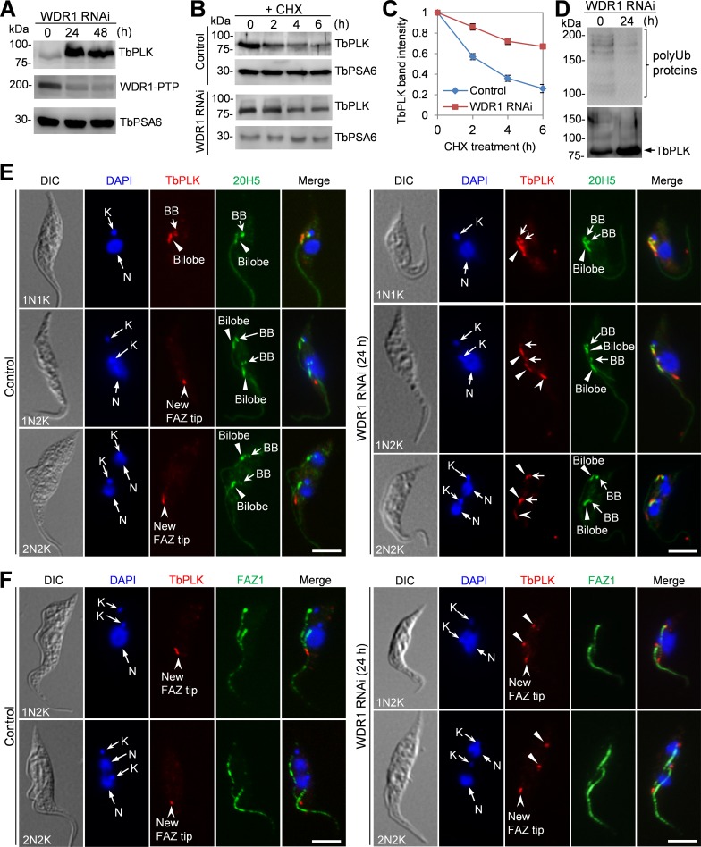
Fig 6. Depletion of WDR1 caused excessive accumulation of TbPLK in the basal body and the bilobe.
(A). Western blotting to detect the level of TbPLK in control and WDR1 RNAi cells that were induced with tetracycline for 24 and 48 hours. TbPLK was detected by anti-TbPLK antibody, and WDR1 was endogenously tagged with a PTP epitope and detected with anti-Protein A antibody. TbPSA6 served as the loading control. Three repeats were performed, and all showed similar results. (B). Degradation of TbPLK in control and WDR1 RNAi cells. The non-induced control cells and WDR1 RNAi cells (24 h) were treated with cycloheximide for up to 6 h, and time course samples were collected at various times after cycloheximide treatment for Western blotting with anti-TbPLK antibody. TbPSA6 served as the loading control. (C). Quantification of TbPLK band intensity shown in panel B. TbPLK band intensity was determined with ImageJ, and normalized with the band intensity of TbPSA6. Error bars indicate S.D. calculated from three independent experiments. (D). Depletion of WDR1 reduced poly-ubiquitinated proteins of TbPLK immunoprecipitates. TbPLK was immunoprecipitated with anti-TbPLK pAb from non-induced control and WDR1 RNAi cells (24 h), and immunoprecipitated proteins were separated by SDS-PAGE and immunoblotted with anti-ubiquitin mAb and anti-TbPLK pAb to detect poly-ubiquitinated proteins (polyUB proteins) and TbPLK, respectively. Three repeats were performed, which showed similar results. (E, F). Effect of WDR1 depletion on TbPLK localization. WDR1 RNAi was induced for 24 h, and cells were co-immunostained with anti-TbPLK pAb and 20H5, which detects centrins in the basal body and the bilobe (panel E), and with anti-TbPLK pAb and anti-FAZ1 mAb, which labels the FAZ filament (Panel F). The arrows, solid arrowheads and open arrowheads in the TbPLK channel of panel E indicate the localization of TbPLK in the basal body, the bilobe and the new FAZ tip, respectively. The solid arrowheads in the TbPLK channel of panel F indicate the localization of TbPLK in the bilobe and the basal body region. N, nuclear DNA; K, kinetoplast DNA. Scale bars: 5 μm.

