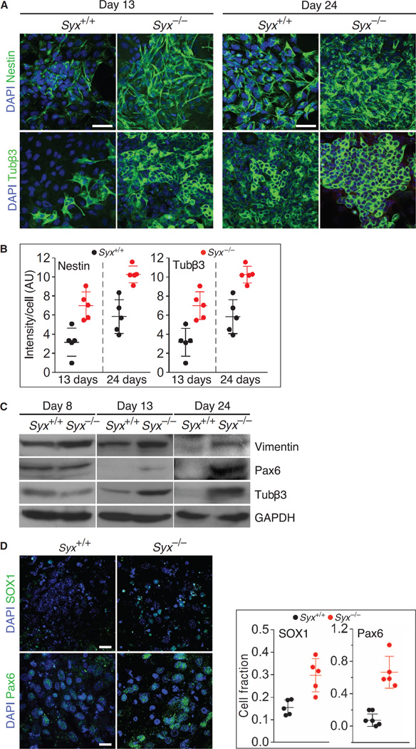
Fig. 1. Neural differentiation in cells from Syx+/+ and Syx−/− EBs.
(A) Immunofluorescence images of nestin- or Tubβ3-labeled cells that were dissociated from RA-treated Syx+/+ and Syx−/− EBs at the indicated times (scale bars, 50 µm). DAPI, 4′,6-diamidino-2-phenylindole. (B) Histograms quantifying nestin and Tubβ3 immunofluorescence intensities shown in (A) [two-way analysis of variance (ANOVA), n = 5 fields, each containing >90 Syx+/+ cells or >130 Syx−/− cells, acquired in two independent experiments; mean ± SD, P < 0.001 for all differences]. AU, arbitrary units. (C) Immunoblot of neural differentiation markers vimentin, Pax6, and Tubβ3 in one of two independent experiments with cells dissociated from RA-treated Syx+/+ and Syx−/− EBs at the indicated time points. GAPDH (glyceraldehyde-3-phosphate dehydrogenase) was used as a loading control. (D) Immunofluorescence images of the neural progenitor cell markers SOX1 and Pax6 in cells from RA-treated Syx+/+ and Syx−/− EBs 12 days after EB aggregation (scale bars, 50 µm). Quantification of the immunofluorescence of each marker is shown in the adjoining histograms [n = 5 fields containing 138 (Pax6, Syx+/+), 82 (Pax6, Syx−/−), 122 (SOX1, Syx+/+), and 93 (SOX1, Syx−/−) cells from two independent experiments; Pax6: 26.2 higher odds for the presence of Pax6 in Syx−/− rather than in Syx+/+ cells (95% confidence interval, 10.6 to 64.8; P < 0.001); SOX1: 2.4 higher odds for the presence of SOX1 in Syx−/− rather than in Syx+/+ cells (95% confidence interval, 1.6 to 3.4; P < 0.001)].

