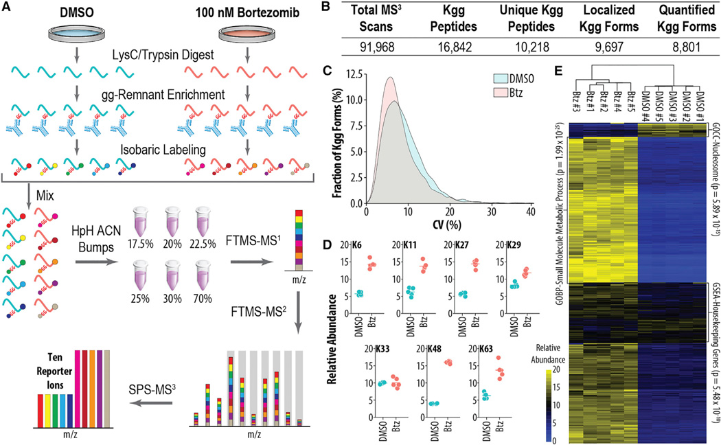
Figure 1. Isobaric Labeling Method for Reproducible, Multiplexed Quantitation of Ubiquitylomes.
(A) Sample preparation and instrumental workflow. Proteins from samples are isolated and digested yielding peptides that are enriched, labeled with isobaric tags, and mixed. Peptides are separated by offline high pH reversed phase (HpH-RP) before analysis by MS using the SPS-MS3 method.
(B) Summary statistics of experiment depicted in (A) are shown.
(C) Coefficient of variation (CV) (n = 5) for either DMSO- or Btz-treated cells is shown.
(D) Quantitative values for ubiquitylated lysine residues on UB are shown.
(E) Global changes in ubiquitylation upon Btz treatment are shown.

