. 2017 Jan 17;6:e22540. doi: 10.7554/eLife.22540

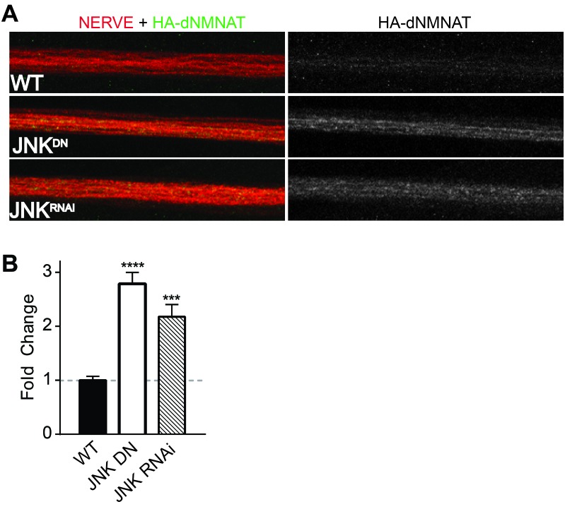
Figure 4. MAPKs modulate dNMNAT in vivo in Drosophila larvae.

Immunostaining in third instar Drosophila larvae of HA-dNMNAT (green) expressed in nerves from the motorneuron driver OK6-Gal4, with HRP labeling (red) to counterstain the nerve. (A) Expression of JNK dominant negative (DN) or depletion of JNK with RNAi (BL57035) increases levels of HA-dNMNAT in the nerve in vivo. (B) Quantification of fluorescence intensity of HA-dNMNAT normalized to wildtype (WT) controls. N = 8–10 animals. p values: ***≤ 0.001, ****≤ 0.0001 by ANOVA. Also refer to Figure 4—figure supplement 1.

DOI: http://dx.doi.org/10.7554/eLife.22540.007

Figure 4.

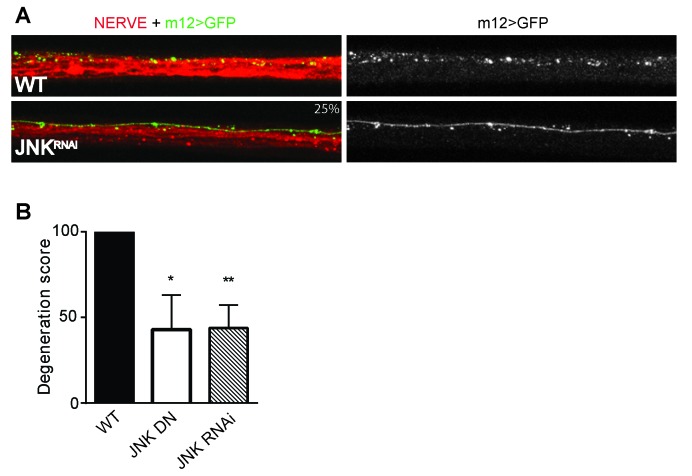
Figure 4—figure supplement 1. Depletion of JNK with RNAi or expression of JNK dominant negative (DN) protects Drosophila larval axons 24 hr after pinch injury.

Figure 4—figure supplement 1.

(A) Representative images of injured axons. Usually two single axons within the nerve are labeled with GFP driven by m12-GAL4. Total nerve is labeled with HRP. (B) Quantification of axon degeneration, with 100 being complete fragmentation and 0 being an intact axon. Values are presented as mean ± SEM. N = 7–12 nerves. **p≤0.01, *p≤0.05.