. 2017 Jan 17;6:e22540. doi: 10.7554/eLife.22540

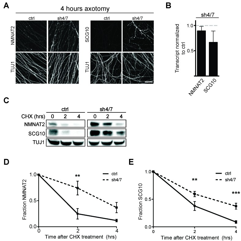
Figure 5. MKK4/7 promote turnover of axon survival factors.

(A) Immunostaining for NMNAT2-myc and SCG10 in distal axons four hours after axotomy. Fluorescence signal is diminished four hours after cut in control axons; however, with depletion of MKK4/7 (sh4/7) levels of NMNAT2-myc and SCG10 are maintained after injury compared to shLuciferase (ctrl) control (p≤0.01 for NMNAT2-myc and p≤0.001 for SCG10, comparing fluorescence between conditions at four hours, n = 4). Scale bar: 25 µm (B) rtPCR reveals no significant change in transcript levels for NMNAT2 or SCG10 after depletion of MKK4/7 when normalized to shLuciferase controls. N = 3, p=0.29. (C) Western blot analysis from axon-only lysate from DRG cultures treated with cycloheximide (CHX) for the indicated timepoints to block protein synthesis. Quantification from CHX timecourse experiments for fraction of NMNAT2 (D) and SCG10 (E) as compared to time zero. The turnover rate of both NMNAT2 and SCG10 is slowed upon MKK4/7 knockdown. N = 4; p values: **≤ 0.01, ***≤ 0.001. Also refer to Figure 5—figure supplements 1 and 2.

DOI: http://dx.doi.org/10.7554/eLife.22540.009

Figure 5.

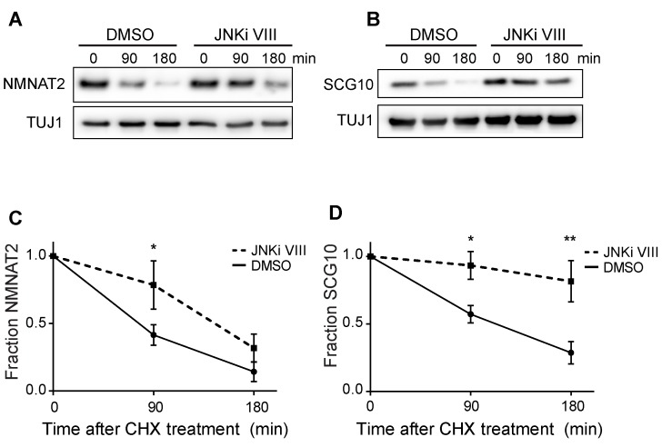
Figure 5—figure supplement 1. Pre-treatment with JNKi VIII (30 min) delays degradation of NMNAT2 and SCG10 after cycloheximide (CHX).

Figure 5—figure supplement 1.

Western blot analysis from axon-only lysate from DRG cultures treated with CHX for the indicated timepoints to block protein synthesis to show turnover of endogenous NMNAT2 (A) and SCG10 (B). TUJ1 is a loading control. Quantification from CHX timecourse experiments for fraction of NMNAT2 (C) and SCG10 (D) as compared to time zero. The turnover rate of both NMNAT2 and SCG10 is slowed upon treatment with JNKi VIII compared to vehicle (DMSO) controls. N = 4; P values: * ≤ 0.05, ** ≤ 0.01.
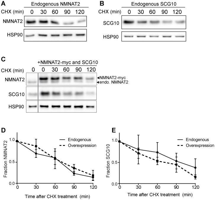
Figure 5—figure supplement 2. Elevation of NMNAT2 and SCG10 does not delay turnover of survival factors within axons.

Figure 5—figure supplement 2.

Representative western blots from axon-only lysate from DRG cultures treated with cycloheximide (CHX) for the indicated timepoints, showing turnover of endogenous NMNAT2 (A) and endogenous SCG10 (B). (C) Representative western blot from axon-only lysate from DRG cultures that were uninfected (left lane; endogenous) or overexpressing both NMNAT2-myc and wildtype SCG10 and treated with CHX. NMNAT2-myc and SCG10 were expressed at levels 5- to 8-fold higher than wildtype protein. The vertical line denotes where bands were cropped out of the blot, but all samples within (C) were run on the same gel. The bands for NMNAT2-myc and endogenous NMNAT2 are labeled. Quantification from CHX timecourse experiments for fraction of NMNAT2 (D) and SCG10 (E) as compared to time zero. The turnover rate of both NMNAT2 and SCG10 is not statistically different when comparing endogenous survival factors in non-infected cultures to the turnover rate in cultures overexpressing high amounts of NMNAT2-myc and SCG10.