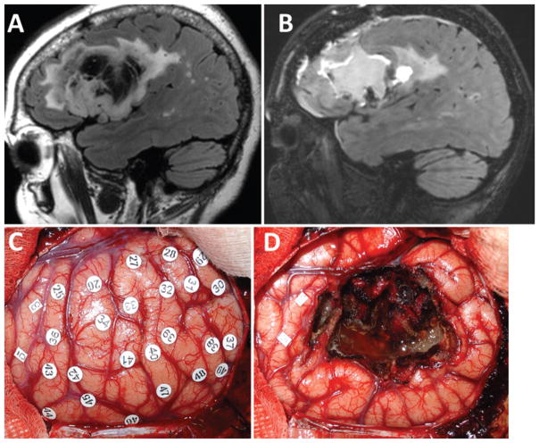
Fig. 3.
Magnetic resonance images (A and B) and intraoperative photographs (C and D) from a representative patient with postoperative language dysfunction. A: Preoperative parasagittal FLAIR image showing the patient’s left frontal opercular oligoastrocytoma involving the pars triangularis and opercularis (but not orbitalis). B: Postoperative FLAIR image showing GTR of the tumor. The patient had transient difficulty with speech initiation postoperatively, which resolved completely within 2 weeks. C: Intraoperative photograph of the operculum prior to resection and after mapping. Stimulation of the area under tag 3 evoked tongue movement, and stimulation of area 5 evoked lip movement. No stimulation produced errors in naming or repetition. D: Intraoperative photograph after the resection was completed showing GTR, confirmed radiologically postoperatively. Figure is available in color online only.

